Quest V-C-M Medium: A Versatile Transport Solution for Diverse Microorganisms
In the realm of clinical microbiology, the efficient and reliable transport of specimens is crucial for accurate diagnoses and timely interventions. Quest V-C-M Medium stands out as a versatile transport solution, effectively preserving and protecting a wide spectrum of microorganisms, including viruses, chlamydiae, mycoplasmas, and now potentially Trichomonas vaginalis.
Preserving the Integrity of Microorganisms
Quest V-C-M Medium's formulation is meticulously designed to safeguard the viability of delicate microorganisms during transport. The inclusion of protein for stabilization ensures that the microorganisms remain metabolically active, minimizing the risk of damage or degradation. Additionally, the medium's carefully balanced composition maintains a neutral pH, an essential factor for the survival of many microorganisms.
Broad-Spectrum Protection Against Contamination
Quest V-C-M Medium incorporates a blend of antibiotics to effectively combat bacterial and fungal contaminants, preventing their overgrowth and ensuring that the target microorganisms remain uncompromised. This protection is particularly crucial for Trichomonas vaginalis, as overgrowth of other microorganisms could interfere with its detection and identification.
Enhancing Coinfection Research
The ability of Quest V-C-M Medium to preserve and protect multiple microorganisms holds significant value for coinfection research. By utilizing a single transport medium, researchers can efficiently collect and analyze samples for multiple pathogens, facilitating a comprehensive understanding of coinfection dynamics and their impact on patient outcomes.
Unlocking the Potential of Trichomonas vaginalis Research
With its demonstrated efficacy in preserving other microorganisms and its suitability for coinfection research, Quest V-C-M Medium emerges as a promising candidate for transporting Trichomonas vaginalis. Further studies to confirm its effectiveness in this application could pave the way for improved diagnostics and treatment strategies for trichomoniasis.
Quest V-C-M Medium: A Cornerstone of Microbiological Research
Quest V-C-M Medium's versatility and effectiveness in preserving a wide range of microorganisms make it an invaluable tool for clinical microbiology. With its potential to extend its protective capabilities to Trichomonas vaginalis, Quest V-C-M Medium continues to evolve as a cornerstone of microbiological research, facilitating accurate diagnoses, furthering our understanding of coinfections, and ultimately improving patient care.
Product Information
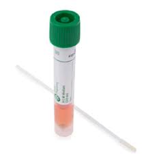
- Product Name: Quest V-C-M Medium 3 mL Vial, carton of 300
- Product Number: 220223
- Manufacturer: Quest Diagnostics
Product Description
Quest V-C-M Medium is a transport medium used to preserve and protect viruses, chlamydiae, and mycoplasmas during transport from the collection site to the laboratory for testing. The medium contains protein for stabilization, antibiotics to minimize bacterial and fungal contamination, and a buffer to maintain a neutral pH.
Features
- Preserves and protects viruses, chlamydiae, and mycoplasmas during transport
- Contains protein for stabilization
- Contains antibiotics to minimize bacterial and fungal contamination
- Contains a buffer to maintain a neutral pH
Applications
- Transport of viral, chlamydial, and mycoplasmal specimens from the collection site to the laboratory for testing
- Use in conjunction with other diagnostic tests for viruses, chlamydiae, and mycoplasmas
Storage
- Store at 2-8°C.
Intended Use: Quest V-C-M Medium is designed for the collection and conveyance of clinical specimens containing viruses, chlamydiae, mycoplasmas, or ureaplasmas from the collection site to the testing laboratory. This system can undergo processing using standard clinical laboratory procedures for viral, chlamydial, mycoplasmal, and ureaplasmal cultures.
Summary and Explanation: In the diagnostic process for infections caused by viruses, chlamydiae, mycoplasmas, or ureaplasmas, the safe and effective collection and transportation of biological samples are crucial. The Quest V-C-M Medium facilitates this process by providing a universal transporting medium that remains stable at room temperature, preserving the viability (and infectivity) of various organisms, including clinically significant viruses, chlamydiae, mycoplasmas, and ureaplasmas during transit to the testing laboratory. The formulation of Quest V-C-M Medium includes proteins for stabilization, antibiotics to minimize bacterial and fungal contamination, and a buffer to maintain a neutral pH.
Quest V-C-M Medium comes with labeled capture-cap vials specifically designed for transporting clinical samples. The capture-cap securely holds the swab sample to the cap, eliminating the need for forceps in the laboratory.
Principles of the Procedure: Quest V-C-M Medium comprises modified Hank’s balanced salt solution supplemented with bovine serum albumin, cysteine, gelatin, sucrose, and glutamic acid. The pH is buffered with HEPES buffer, with Phenol red indicating pH levels. Vancomycin, amphotericin B, and colistin are incorporated into the medium to inhibit the growth of competing bacteria and yeast. The medium is isotonic and non-toxic to mammalian host cells. The presence of sucrose serves as a cryoprotectant, aiding in the preservation of viruses and chlamydiae if specimens are frozen (–70°C) for extended storage.
Specimen Collection and Preparation:
For investigations involving viruses, chlamydiae, mycoplasmas, or ureaplasmas, specimens should adhere to established manuals and guidelines. To ensure optimal viability, promptly transport specimens to the laboratory. Refrigeration at 2–8°C or placement on wet ice during transit is recommended, while prolonged delays necessitate freezing at –70°C or colder, transported on dry ice. Adherence to state and federal regulations for specimen shipment and handling is imperative, with internal institution guidelines applicable for in-house specimen transfers. Process all received specimens promptly upon arrival at the laboratory.
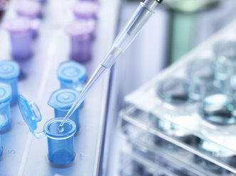
Procedures: Materials Provided: Quest V-C-M Medium includes a capture-cap vial containing 3 mL of transport medium plus three glass beads. Materials Required But Not Provided: Specimen collection swab, materials for isolating, differentiating, and culturing viruses, chlamydiae, mycoplasmas, and ureaplasmas.
Test Procedure:
- Aseptically remove the cap from the vial.
- Aseptically place the sample into the vial with medium.
- Replace the cap on the vial and close tightly.
- Label with appropriate patient information.
- Send to the laboratory for immediate analysis.
Quality Control: All lots of V-C-M Medium undergo testing for microbial contamination, toxicity to host cells, and the ability to maintain viability. Non-compliance with quality control standards warrants withholding patient results.
Results: The obtained results are contingent upon proper specimen collection, timely transport, and laboratory processing.
Limitations of the Procedure:
- Variables like the condition, timing, and volume of specimens collected for culture significantly impact reliable results. Adherence to recommended guidelines for specimen collection is crucial.
- Repeated freezing and thawing may reduce recovery of viable organisms.
- V-C-M Medium is intended for collection and transport of viral, chlamydial, mycoplasmal, and ureaplasmal agents. It also serves as a cryoprotectant for clinical viruses.
- Specific swab materials should be used to avoid interference with tests, and the product's performance is validated with BD™ Universal Viral Transport Swabs only.
Performance Characteristics:
Quest V-C-M Medium, validated with BD™ Universal Viral Transport Swabs, demonstrated viability maintenance for various organisms up to 48 hours at both room temperature (20–25°C) and refrigerated conditions (2–8°C). Tested organisms include adenovirus, cytomegalovirus, echovirus type 30, herpes simplex viruses, influenza A, parainfluenza 3, respiratory syncytial virus, varicella-zoster virus, Chlamydophila pneumoniae, Chlamydia trachomatis, Mycoplasma hominis, Mycoplasma pneumoniae, and Ureaplasma urealyticum.
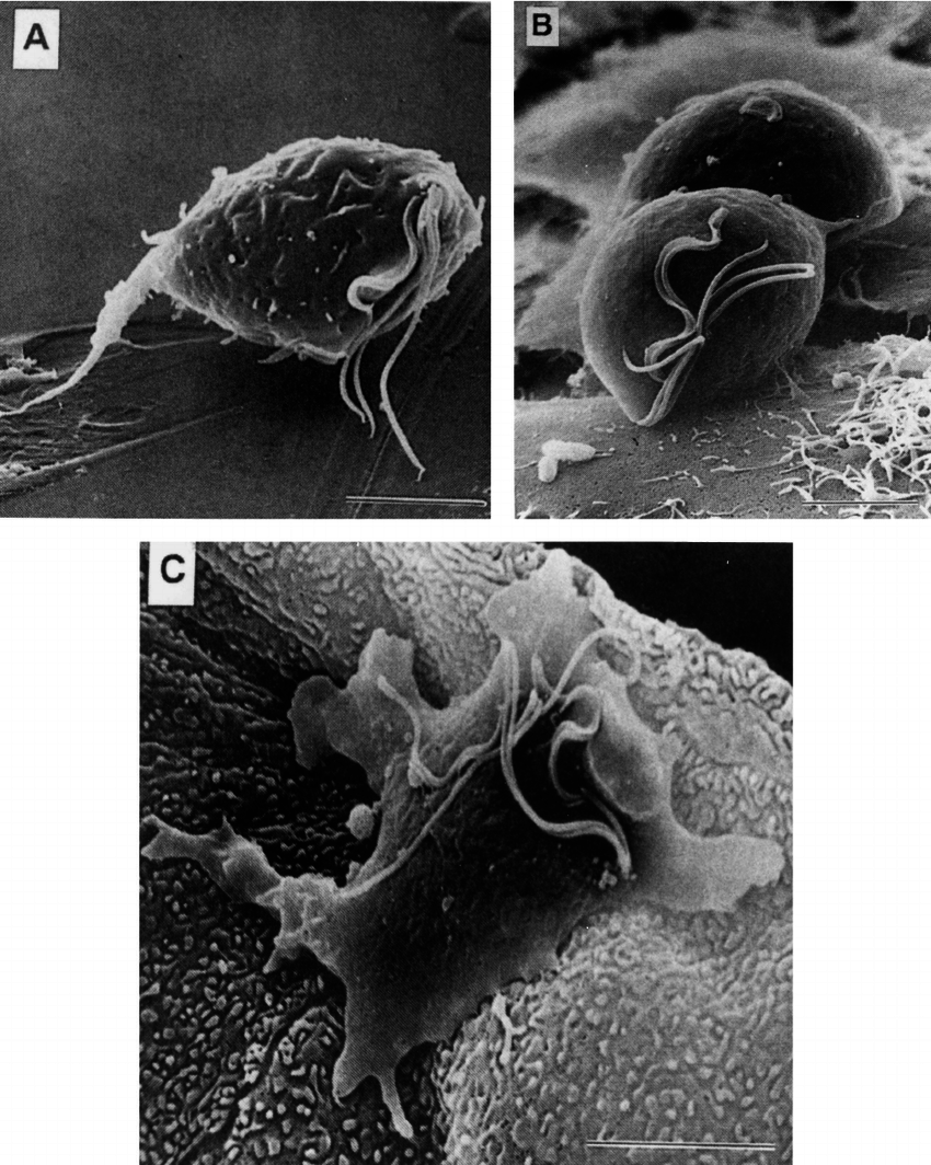
Microscopic Exploration of Trichomonas vaginalis: Unveiling Unique Morphological Characteristics
Delving into the microscopic world of Trichomonas vaginalis reveals a fascinating array of morphological features that define this parasitic protozoan. Through the lens of a microscope, these pear-shaped organisms emerge with their distinctive features, providing valuable insights into their biology and pathogenic potential.
Pear-Shaped Silhouette: A Hallmark of Trichomonas vaginalis
The most striking characteristic of Trichomonas vaginalis is its distinctive pear-shaped morphology. This elongated shape, with a rounded anterior end and a tapering posterior end, sets it apart from other microorganisms. The pear-shaped silhouette facilitates the parasite's movement within the genitourinary tract, enabling it to navigate through mucosal surfaces and evade host defenses.
Flagellar Ensemble: Propulsion and Sensory Guidance
Adorning the anterior end of Trichomonas vaginalis is a cluster of four flagella, each serving a specific function. Three of these flagella are responsible for propulsion, enabling the parasite to glide and rotate through its environment. The fourth flagellum, known as the recurrent flagellum, extends along the body and plays a sensory role, aiding in chemotaxis and attachment to host cells.
Undulating Membrane: A Dynamic Outer Covering
The surface of Trichomonas vaginalis is characterized by an undulating membrane, a dynamic structure that constantly folds and unfolds. This undulating motion not only contributes to the parasite's motility but also enhances its adherence to host cells. The membrane also plays a role in nutrient uptake and evasion of host immune responses.
Unique Morphological Features: A Reflection of Parasitic Adaptations
The combination of unique morphological features, including its pear-shaped silhouette, flagellar ensemble, and undulating membrane, equips Trichomonas vaginalis with remarkable adaptations for parasitism. These features enable efficient movement within the host environment, facilitate attachment to host cells, and contribute to nutrient acquisition and immune evasion.
Unveiling the Microscopic World of Trichomonas vaginalis: A Window into Pathogenicity
The microscopic examination of Trichomonas vaginalis provides valuable insights into its morphology, revealing adaptations that contribute to its parasitic lifestyle. By understanding these unique features, we can gain a deeper understanding of the parasite's pathogenic mechanisms and develop effective strategies for prevention and treatment.